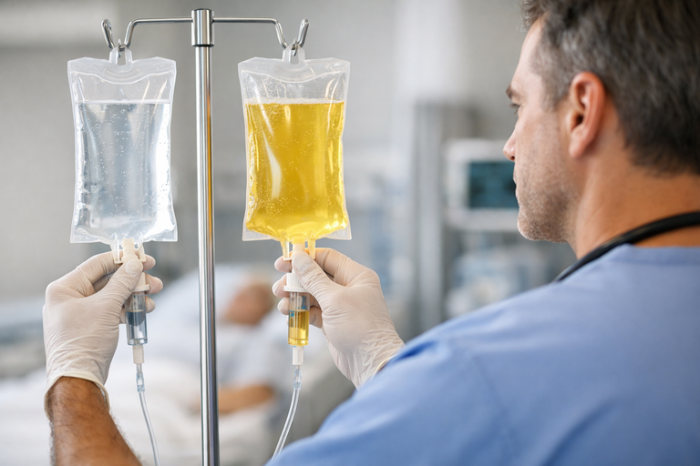
When most of us imagine medical treatment, we think of operating rooms, long incisions, and lengthy recoveries. But medicine has quietly shifted in a different direction—one where tiny tools, live imaging, and precise navigation can treat major health problems without traditional surgery. This field is known as Interventional Radiology, and it has transformed care for millions of patients around the world.
In this post, we’ll explore how it works, why it’s becoming so important, and what happens behind the scenes with the specialists and technologies that make it possible.
What Exactly Is Interventional Radiology?
At its core, interventional radiology uses imaging—X-ray, ultrasound, CT, and sometimes MRI—to guide miniature instruments through a pin-sized opening in the skin. Instead of cutting open the body, doctors navigate catheters, wires, or needles to the exact location of a problem.
Think of it as treating conditions from the inside out.
With this approach, patients often experience:
- Smaller entry points instead of surgical cuts
- Lower risk of infection
- Less pain and less anesthesia
- Faster return to daily life
Interventional radiology has become a quietly powerful option for treating everything from blood vessel disease to cancers, organ blockages, and even painful spinal fractures.
If you’re curious about how these advanced methods work in practice, you can explore the full overview here: Interventional Radiology.
The Technology Behind the Precision
Modern procedures depend on highly skilled teams and sophisticated tools. This is where interventional radiology tech plays a vital role.
Interventional radiology technologists assist with:
- Operating imaging equipment during procedures
- Preparing tools such as microcatheters and guidewires
- Ensuring sterile protocol
- Monitoring radiation safety
They act as the bridge between the radiologist, the patient, and the technology—making sure every movement is accurate down to the millimeter.
What Conditions Can Be Treated?
The surprising answer: many conditions once treated with open surgery.
1. Vascular Disease
This is where vascular interventional radiology shines. Blood vessel problems—arteries or veins—can often be repaired from within.
Examples include:
- Peripheral arterial disease (PAD): Opening narrowed leg arteries to improve walking and wound healing
- Deep vein thrombosis (DVT): Removing or dissolving clots in selected cases
- Pulmonary embolism (PE): Clearing dangerous clots from lung vessels
- Varicose veins: Closing abnormal veins that cause heaviness, swelling, and discomfort
These procedures generally require only local anesthesia and allow patients to walk out the same day.
2. Cancer and Tumor Care
Interventional oncology is one of the fastest-growing areas in the field.
Techniques include:
- Ablation: Destroying tumors using heat, cold, or electric pulses
- Embolization: Cutting off blood supply to tumors
- Liver-directed therapies: Targeting cancers that cannot be removed surgically
These are highly targeted treatments focused on tumor control while preserving healthy tissue.
3. Organ Blockages and Drainage
When bile ducts, kidneys, or other organs become blocked, interventional radiologists can place stents or drains to relieve pressure and restore function.
Procedures include:
- Biliary drainage
- Nephrostomy
- Abscess or fluid drainage in the abdomen, chest, or soft tissues
These small interventions can rapidly improve pain, infection, and overall health.
4. Spine and Pain Relief
For patients with vertebral compression fractures—often due to osteoporosis—procedures such as vertebroplasty or kyphoplasty reinforce the weakened bone and reduce pain almost immediately.
What to Expect Before, During, and After a ProcedureBefore
Patients undergo evaluation, blood tests, and imaging. Instructions regarding fasting, medication adjustments, and allergies (especially to contrast dye) are provided.
During
Most procedures use local anesthesia with light sedation.
Real-time imaging guides the physician as they move through tiny blood vessels or tissues.
After
Recovery is usually quick—often just a few hours of monitoring. Many people return to normal routines within 24–72 hours.
Neuro Interventional Radiology: A Special Frontier
One of the most dramatic uses of these techniques is in the brain.
Specialists can now:
- Remove clots causing acute stroke through mechanical thrombectomy
- Treat brain aneurysms using coils or flow-diverting stents
- Widen narrowed carotid arteries with stents
In stroke care, every minute matters—procedures done quickly can significantly improve outcomes.
Why It Matters
Interventional radiology represents a shift in modern medicine: away from large incisions and long hospital stays, toward precise, image-guided solutions that preserve quality of life.
It’s not only about convenience—it’s about giving patients safer alternatives, especially when surgery isn’t ideal.
Final Thoughts
Whether it’s restoring blood flow in the legs, treating a tumor, stopping internal bleeding, or relieving a painful blockage, interventional radiology has reshaped what minimally invasive care looks like.
If you’ve ever wondered how medical teams navigate tiny vessels deep inside the body, or how life-saving treatments can happen through just a few millimeters of skin, this specialty is worth exploring.
For a closer look at techniques, conditions, and expert teams, you can visit the full resource here:👉 Interventional Radiology